| Back Futurehealth | |||||||
|
Original Content at https://www.futurehealth.org/articles/Neurofeedback-Treatment-of-by-Paul-Swingle-090324-767.html |
|||||||
March 24, 2009
Neurofeedback Treatment of Pseudoseizure Disorder
By Paul Swingle
Background: Previous research has shown that the suppression of theta wave activity and the enhancement of Sensorimotor Rhythm (SMR) through electroencephalographic (EEG) biofeedback is an effective treatment for epilepsy. The current research reports the results of EEG biofeedback treatment for patients presenting with seizure behaviors in the absence of epileptiform EEG activity.
::::::::
Background: Previous research has shown that the suppression of theta wave activity and the enhancement of Sensorimotor Rhythm (SMR) through electroencephalographic (EEG) biofeedback is an effective treatment for epilepsy. The current research reports the results of EEG biofeedback treatment for patients presenting with seizure behaviors in the absence of epileptiform EEG activity.
Methods: In addition to psychotherapy, 3 patients, 2 women and 1 man, were trained, using EEG feedback once per week, to reduce the ratio of theta band to SMR band EEG amplitudes.
Results: The results showed that reductions in seizure activity were related to reductions in the theta – SMR ratio.
Conclusions: These findings support the view that theta – SMR feedback training, in conjunction with psychotherapy, is an effective adjunctive treatment for pseudoseizure disorder. Biological Psychiatry 1998; 44(11); 1196-1199. © 1998 Society of Biological Psychiatry
Key Words: Neurotherapy, electroencephalographic feedback, pseudoseizures, nonepileptic events, biofeedback
Introduction
Pseudoseizures are sudden changes in behavior that appear to be seizures but without any identifiable organic cause (Bowman and Markand 1996). Such nonepileptic events (NEE) may be characterized by epileptic-like seizures, numbness in the face and extremities, fainting, and fugue-like states. Because pseudoseizure patients manifest high levels of psychiatric comorbidity (Lempert and Schmidt 1990; Bowman 1993) and no identifiable neurological anomalies, it is assumed by many clinicians that such NEE are psychological in nature (Bowman and Markand 1996).
Pseudoseizure disorder is puzzling because of the lack of significant EEG anomalies. Such patients often
From the School of Psychiatry, Ottawa University, Ottawa, Canada.
Address reprint requests to Paul G. Swingle, PhD, School of
Psychology, Ottawa University, Ottawa, K1N 6N5, Canada.
Received November 4, 1996; revised August 27, 1997; accepted
September 10, 1997
![]() © 1998 Society of Biological Psychiatry
© 1998 Society of Biological Psychiatry
have epileptic histories together with a history of traumatic abuse. Hence, one hypothesis regarding pseudoseizure disorder is that the patient has learned, from past epileptic seizures, behaviors that serve to mask abreactive breakthroughs of past trauma.
An alternative to the above conceptualization is to view NEE as essentially an arousal disorder. Such a view is not inconsistent with the “breakthrough” hypothesis, but rather conceptualizes the NEE as being triggered by stimuli that intensify autonomic arousal. Such arousal could be associated with environmental stimuli such as loud noises, stimulants such as caffeine, or emotional content such as traumatic memories.
This report describes the use of neurofeedback therapy to reduce the electroencephalographic (EEG) theta/sensorimotor rhythm (SMR) ratio for patients who present with NEE. The effectiveness of neurofeedback therapy to reduce theta band (4-7 Hz) activity and enhance SMR (12-14 Hz) activity, as measured at location Cz (International 10/20 system), in the treatment of epilepsy is documented (Sterman 1986), although no data are available for pseudoseizure patients. The case studies to be reported below are consistent with the population of patients reported by Bowman and Markand (1996). All had long histories of therapeutic intervention with limited success. Further, EEG assessments showed no epileptiform activity before, during, or following a seizure. All presented with significant comorbidity and poorly explained seizure onset in adulthood, and all have had multiple psychiatric hospitalization.
A treatment protocol was developed to modify the theta/SMR ratio, which included brainwave disentrainment (Rozelle and Burzynski 1995) and neurofeedback (Ayers 1993). Brainwave disentrainment involves measuring the dominant EEG frequency (DF) (i.e., maximum amplitude averaged over 1-sec epochs) and stimulating the client with light-emitting diode goggles and sounds at a frequency either 5% greater than or 5% less than the DF. These multipliers (i.e., + or – 5%) alternate every minute, with the result that the brain’s DF extends over a wider range. The purpose of this procedure is to enhance neuronal flexibility and normalize the EEG (Tachiki et al 1994). Positive results from the procedure have been reported for the treatment for mild head injury (Ochs 1994) and learning disability (Russell et al 1994).
0006-3223/98/$19.00
PII S0006-3223(97)00541-6
SCL
(μMHO)
Figure 1. Skin conductivity level (SCL) during auditory stimulation of a client with pseudoseizure disorder.
The neurofeedback protocol is based on the work of Sterman (1986) and Ayers (1993). It involves the suppression of the theta band activity concordantly with the enhancement of SMR band activity. Neuronal feedback to the patient occurred when SMR band activity exceeded a set threshold and when theta band activity was below a set threshold. A monopolar montage was used with the active electrode at Cz (International 10/20 system). Frequency of treatment was between one and three times per week. Notably, all were being treated concurrently with psychotherapy during the adjunctive theta/SMR training.
Case One
The patient, a Caucasian woman in her early forties, experiences a variety of seizure-like episodes that occur with normal waking EEG architecture. The patient has identified several NEE subtypes, including fugue-like states, a loss or compromise of motor and/or cognitive control, and episodes of rage. The severe NEE typically involve collapsing to the floor with the body assuming a fetal position. In that position the torso convulses and the arms and/or legs may thrash about. Typically these severe NEE last from several seconds to several minutes and may occur repeatedly. This patient has been hospitalized for various psychiatric complaints over 10 times during the last 10 years. This patient has a long and significant history of severe physical , psychological, and sexual abuse. This patient was intolerant of light and sound stimulation, a condition often observed in posttraumatic stress disorder patients. Her tolerance of various sounds was assessed while electrodermal activity (EDR) was measured, from both hands, using a constant voltage electrodermal monitor. As noted in Figure 1, seizures occurred when the EDR was greater than about 4 mho in either hand. A procedure for reducing autonomic arousal (Swingle 1994), which includes the presentation of subthreshold sounds and/or somatosensory stimulation, was presented during the auditory stimulation.
SCL
(μMHO)
Figure 2. Skin conductivity level (SCL) during auditory stimulation of a client with pseudoseizure disorder.
Figure 2 shows the EDR level for one session in which arousal was reduced while sound stimulation was presented. The protocol maintained the EDR below 4 mho, and no seizures were observed.
The outcome data, separated into four categories, are from very detailed records maintained by the patient at the suggestion of her neuroendocrinologist in September of 1993. The four categories are the total number of seizures, SFD, MS (3 or more per day), and SS. After 11 months of treatment (modal frequency once per week) SFD increased by 15%, MS decreased by 8%, and the total number of seizures per month decreased by 30% (t < 2.5, df = 8, p < .05 in all cases).
The patient was able to modify her theta/SMR ratio during the disentrainment and neurofeedback treatment. The average theta/SMR ratio recorded during pretreatment baseline on each day of treatment for the first 3 months (mean = 1.96, SD = 0.12) and for the last 3 months of treatment (mean = 1.55, SD – 0.20) were found to be significantly different (t = 3.55, df = 23, p < .01). As the data in Table 1 indicate, the theta/SMR ratio correlates with SS and the total number of seizures, marginally with MS, but not with SFD.
The daily seizure totals were also compared with the theta/SMR average on the days of EEG treatment. The theta/SMR ratio for days when no seizures were reported (mean = 1.46, SD = 0.113) differed significantly (t = 2.87, df = 28, p < .02) from seizure days (mean = 1.68, SD = 0.228).
Table 1. Spearman Rank-Order Correlations between Theta/SMR ratio and categories of Reported Seizure Activity
Category of seizure activity Coefficient p
Multiple-seizure days .56 .07
Severe seizures .75 .05
Seizure-free days .07 þu
Total seizures .78 .05
The data indicate that the theta/SMR ratio is associated with the self-rated seizure activity, and that the treatment protocol resulted in a significant deduction in the theta/SMR ratio.
Case Two
The second patient, a Caucasian man in his early forties, was referred for treatment of “paranoid/panic” attacks. This patient complained that his hands would tremble, and he would become frightened, perspire heavily, experience a numbing of the left side of his body, and feel as though a policeman was standing close to his left side.
The referring psychopharmacologist had required the patient to maintain a record of the attacks, which indicated that for the month prior to neurotherapy treatment, the patient experienced 16% attack-free days (AFD) and 42% multiple-attack days (MAD) (two or more attacks). Although EEG evaluations did not reveal any anomalous activity, this patient did manifest high theta amplitudes as measured at Cz with theta/SMR ratios in the 4 – 5 range (mean = 4.88, DF = 1.32). Treatment consisted of theta suppression and SMR enhancement at Cz as described in case one. The results indicate that the patient’s attacks were related to the theta/SMR ratio (r = .60, df = 26, p < .01, two tailed).
Case Three
The third case, a Caucasian woman in her mid sixties, was referred for treatment of “fainting spells.” During these spells, the patient would appear to fall asleep, often falling from her chair. Her right arm and her head would shake rhythmically during some of these spells. This patient had multiple hospitalizations for conditions other than her pseudoseizures including obsessive-compulsive disorder and depression.
Initial EEG baseline indicated a theta/SMR ratio of 1.5. During the course of the initial assessment the patient experienced a fainting spell (there were no hand or head movements during this episode), during which time the theta/SMR ratio averaged 3.64.
The treatment for this patient included EEG disentrainment and theta/SMR neurofeedback. Because this patient experienced seizures in session, the relationship between the theta/SMR ratio and her fainting spells could be determined. These data are shown in Figure 3.
The pretreatment theta/SMR ratio on days when the patient experienced a fainting spell during treatment was not different from a seizure-free session but was significantly larger when the patient reported
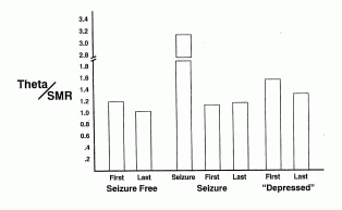
Figure 3. Two-minute baseline recording of the average that/SMR ratio at the beginning and end of neurotherapy sessions with a client with pseudoseizure disorder. Measures took place during client-reported seizure-free, seizure, and depressed periods. The theta/SMR ratios during an in situ seizure are presented with the data collected from the seizure period.
“depression” (t = 2.87, df = 12, p < .02). The theta/SMR ratio during a seizure was significantly different from all the other averages (p < .01, two tailed in all cases). The seizures remitted during the third week of treatment, when the theta/SMR ratio stabilized at an average of 1.06 (SD = 0.12).
Discussion
Lubar and Bahler (1976) have reported that if high amplitude slow-wave activity occurs in conjunction with SMR activity, either a transient increase in seizure rate or a lack of seizure decrease occurs with epileptic patients. This reported finding is consistent with the findings reported here indicating a relationship between the theta/SMR ratio and seizure activity. Further, the observation that seizure activity of the patient described in case one was also associated with her electrodermal activity suggests that pseudoseizure disorder may be essentially an arousal disorder. The beneficial effects of SMR training may be that the SMR inhibits or suppresses excitation in the sensorimotor area.
In summary, although psychological mechanisms appear prevalent in pseudoseizure disorder, and some patients experience reductions or elimination of seizures during the first year after diagnosis, the cases reported here had all been diagnosed a minimum of 7 years prior to theta/SMR treatment. It is apparent, therefore, that seizure activity is directly related to the theta/SMR ratio, and that reductions in the theta/SMR ratio brought about by neurofeedback are associated with reductions in seizure behavior. Due to the rare nature of this disorder, however, control groups are difficult to obtain, which in turn limits the extent of these findings. Nonetheless, the use of theta/SMR
training to reduce seizure behavior concurrently with psychotherapy to address the contributing psychological issues seems prudent to facilitate enhanced social functioning of patients with pseudoseizure disorder.
Portions of the data from the first case report were presented at the meetings of the Society for the Study of Neuronal Regulation, Las Vegas, Nevada, 1994.
References
Ayers ME (1993): A controlled study of EEG neurofeedback training and clinical psychotherapy for right hemisphere closed head injury [abstract]. In: Proceedings of the Association for Applied Psychophysiology and Biofeedback. pp 19-20.
Bowman ES (1993): Etiology and clinical course of pseudoseizures: Relationship to trauma, depression, and dissociation. Psychosomatics 34:333-342.
Bowman ES, Markand ON (1996).: Psychodynamics and psychiatric diagnoses of pseudoseizure subjects. American Journal of Psychiatry, 153:57-63
Hoffman DA, Stockdale S, Hicks LL, Schwaninger JE (1995): Diagnosis and treatment of head injury. Journal of Neurotherapy, 1:14-21.
Lempert T, Schmidt E (1990): Natural history and outcome of psychogenic seizures: A clinical study in 50 patients. Journal of Neurology, 237:35-38.
Lubar JF, Bahler WW (1967): Behavioural management of epileptic seizures following biofeedback training of the sensorimotor rhythm. Biofeedback and Self-Regulation, 1:77-104.
Lubar JF, Shouse MN (1976): EEG and behavioural changes in a hyperkinetic child concurrent with training of the sensorimotor rhythm (SMR): A preliminary report. Biofeedback Self Regulation, 3:293-306.
Mosmans PCM, Jonkman EF, Magnus O, Van Huffelen AC (1973): Regional cerebral blood flow and EEG. EEG and Clinical Neurophysiology, 33:122.
Ochs L (1994): EEG-driven stimulation and heterogeneous mild head injured patients: Extended observations. Paper presented at the 2nd Annual Conference of the Society for the Study of Neuronal Regulation, Las Vegas, Nevada.
Roy A (1979): Hysterical seizures. Archives of Neurology, 36:447.
Rozelle GR, Burzynski TH (1995): Neurotherapy for stroke rehabilitation: A single case study. Biofeedback and Self-Regulation, 20:211-228.
Russell HL, Carter JL, Ochs L (1994): Use of EEG/stimulation training with learning disabled (LD) boys: A controlled study. Paper presented at the 2nd Annual Conference of the Society for the Study of Neuronal Regulation, Las Vegas, Nevada.
Sterman MB (1986): Epilepsy and its treatment with EEG feedback therapy. Annuals of Behavioral Medicine, 8:21-25.
Sterman MB, Friar L (1972): Suppression of seizures in an epileptic following sensorimotor EEG feedback training. Electroencephalography and Clinical Neurophysiology, 33:89-95.
Sterman MB, MacDonald LR, Stone RK (1974): Biofeedback training of the sensorimotor EEG rhythm in man: Effects on epilepsy. Epilepsia, 15:395-417.
Swingle PG (1994): Brainwave treatment of pseudoseizure disorder. Paper presented at the 2nd Annual Conference of the Society for the Study of Neuronal Regulation, Las Vegas, Nevada.
Tachiki KH, Ochs L, Weiler E (1994): Brain entrainment as a tool in brainwave retraining. Paper presented at the 2nd Annual Conference of the Society for the Study of Neuronal Regulation, Las Vegas, Nevada.
Tansey MA, Bruner RL (1983): EMG and EEG biofeedback training in the treatment of a 10-year-old hyperactive boy with a developmental reading disorder. Biofeedback Self-Regulation, 8:25-37.
Authors Bio:
Paul G. Swingle, Ph.D. was Professor of Psychology at the University of Ottawa prior to moving to Vancouver. A Fellow of the Canadian psychological Association, Dr. Swingle was Lecturer in Psychiatry at Harvard Medical School from 1991 to 1998 and during the same time period was Associate Attending Psychologist at McLean Hospital where he was also Coordinator of the Clinical Psychophysiology Service. Dr. Swingle was Chairman of the Faculty of Child Psychology at Ottawa University from 1972 to 1977 and Clinical Supervisor from 1987 to 1997. He is a Registered Psychologist in British Columbia and is Certified in Biofeedback and neurotherapy.