| Back Futurehealth | |||||||
|
Original Content at https://www.futurehealth.org/articles/Oral-Pharyngeal-Dysphagia-by-Maggie-Lee-Huckabe-091023-368.html |
|||||||
October 23, 2009
Oral Pharyngeal Dysphagia; Application of EMG Biofeedback in the Treatment of Oral Pharyngeal Dysphagia
By Maggie Lee Huckabee, M.A., CCC/SLP
EMG biofeedback presents auditory and visual signals to aid the patient in learning and practicing dysphagia intervention exercise. Biofeedback provides several critical elements that are unavailable with standard dysphagia therapy.
::::::::
IntroductionOral-pharyngeal dysphagia is a common sequelae to a variety of acute and chronic illnesses, including cerebrovascular accident(16), traumatic brain injury(9), oral-pharyngeal carcinoma(2) and a myriad of degenerative neuromuscular diseases(5).
Swallowing impairment can result in significantly delayed recovery, malnutrition and dehydration(14), pulmonary complications leading to aspiration pneumonia and potentially death(3). Common deficits include weakness in the musculature of the lips, mouth, tongue, palate, pharynx and esophagus, incoordination of movements, surgical ablation of tissue and decreased sensation of the oral and pharyngeal cavities leading to a delay in the stimulation of the swallow(11).
In the past 5-10 years, there has been a significant movement toward rehabilitative treatment of dysphagia. Dysphagia intervention is utilized to both compensate for oral-pharyngeal swallowing impairment and to change the physiology of the swallow through muscle retraining, thus resulting in long term improvement in swallowing(11).
Many of the identified treatment strategies require that the patient translate a previously automatic physiological function into a volitionally controlled, altered motor response. For many patients, both those with and without associated cognitive impairment, grasping the concept of altering swallowing behavior is a challenge. There is little cognitive awareness as to how a swallow is executed. The swallowing therapist is consequently often confronted with confusion, frustration and an inability of the patient to participate in his or her own treatment.
EMG biofeedback presents auditory and visual signals to aid the patient in learning and practicing dysphagia intervention exercises(4). The EMG signal translates the previously automatic response into a concrete representation of oral-pharyngeal movement. Viewing the swallow as it occurs allows the patient to consciously monitor and alter swallowing behavior. The patient is able to clearly visualize the effects of specific compensatory and therapeutic strategies and target visible, realistically attainable goals. Additionally, in times of restrictions by third-party payers, EMG biofeedback provides quantitative data to document patient progress with treatment.
Treatment Protocol
Treatment of oral-pharyngeal swallowing impairment relies on accurate diagnosis. Thus any treatment protocol should be preceded by a thorough diagnostic evaluation by a qualified speech language pathologist. Specific exercises for the treatment of neurophysiological deficits have been described in the dysphagia literature(1,10,11). The scope of this protocol does not include an exhaustive description of dysphagic abnormalities and the associated treatment strategies of choice. It will encompass description of those treatment strategies that are appreciably enhanced by biofeedback monitoring.
Using MyoDac 2TM / MyoCompTM
The MyoComp System, as well as other computer assisted EMG Biofeedback Systems, allows for long-term storage of patient biographical, insurance and medical information on a separate diskette. This is accessed through the database mode on the main menu. In addition, a progress note section enables the therapist to store session information regarding the nature of treatment provided, the patients response to treatment, and other pertinent information. This information can be added to the diskette before or after a session through the monitoring mode on the main menu.
Setting on MyoDac 2TM
Setting on MyoCompTM System
- Options-as desired for auditory feedback and visual
contrast of EMG line on background.
Monitor
- For two channel tasks, use Channel 1 Top, Channel 2 Bottom.
- For one channel tasks, use Channel 1 Center.
- Time-as desired. A setting of 15 seconds provides
more responsive input to patients in the early stages
of treatment.
Single Channel Monitoring
For non-computerized monitoring or home training, the portable MyoTracTM single channel electromyograph can be used.
Oral Motor Facial Exercises
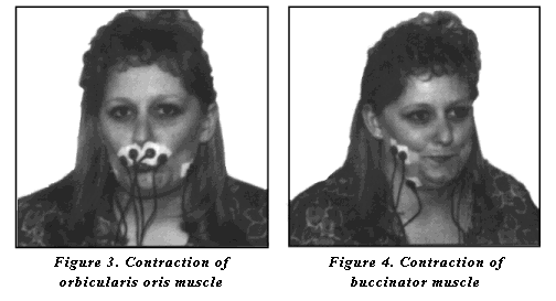
In cases of unilateral cerebrovascular accident or surgical resection and scarring from oral-pharyngeal carcinoma, facial weakness and asymmetry may inhibit adequate bolus control during feeding. Oral motor/facial exercises may be used to improve function in weakened muscles or to reduce constriction associated with scarring. Two muscles have been targeted for retraining with biofeedback. The orbicularis oris is a circular-appearing muscle that extends posteriorly from the buccinator muscle, with upper and lower muscle fibers inserting into the upper and lower lip. This muscle is functionally responsible in the deglutitive process for maintenance of the bolus within the oral cavity. The buccinator muscle is a flat band of muscle that provides the framework for the cheek. This muscle attaches at the pterygomandibular raphe with insertion into the orbicularis oris. The buccinator, combined secondarily with the masseter muscle, serves to tighten the cheeks during mastication and swallowing, inhibiting buccal pocketing. When the goal of treatment is to increase symmetry of contraction of the orbicularis oris and buccinator muscles, as in the case of unilateral cerebral infarcts, biofeedback can be used to assist the patient in matching the weakened musculature to the stronger intact side.
Electrode Placement: The EMG sensors are attached with surface single electrodes, connected to the MyoScan(TM) sensor with electrode extender leads. As functional-facial symmetry is the goal, two channels are used for the patient to target function of weakened hemiparetic side to parallel function of the stronger functioning side. The active electrodes are placed over the orbicularis oris or buccinator muscle with the ground electrode placed anterior to the muscle (figure 1 and 2). For work on lip closure, the function of both the upper and lower lip can be targeted; however, the upper lip is more accessible for secure electrode placement.

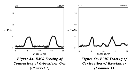
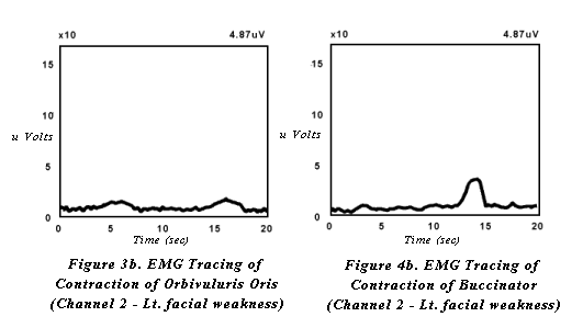
When targeting increased lip strength and closure, the patient is instructed to repetitively purse their lips then relax (figure 3). When targeting increased buccal tension, patients are instructed to tighten the cheeks against the teeth then relax (figure 4). Modeling of the task is usually required. With each contraction of orbicularis oris or the buccinator muscles and surrounding musculature, the patient will see a slope in the receiving, feedback line. The hemiparetic side will demonstrate decreased visual feedback (figures 3a, 3b and 4a, 4b). The goal for treatment is to shape the hemiparetic side to match the stronger, intact side of the face. Intermittent tactile stimulation may be applied by the therapist to increase sensitivity and response between volitional exercise.
Pharyngeal Phase Exercises
Electrode Placement: One triode surface electrode is placed vertically under the chin. The two active electrodes are aligned centrally, spaced evenly between the inferior tip of the mandible and the thyroid cartilage (figure 5). Palpation is required for accurate placement.
Figure 5
Modified Valsalva / Effortful Swallows
A common diagnostic finding in the dysphagic population is decreased or weakened contraction of the pharyngeal constrictors responsible for propulsion of the bolus through the pharynx. Weakness of these muscles results in post-swallow pharyngeal residual of the bolus, placing the patient at risk for post swallow aspiration. A modified valsalva swallow, or hard swallow can be used as a compensatory strategy to aid pharyngeal clearing during a meal and also as an indirect exercise for increasing pharyngeal constriction. The superior, middle and inferior constrictors are internal pharyngeal muscles that are not easily accessible by surface electrode. Direct EMG measurement of these muscles is not feasible. Although further investigation is required to specifically document the correlation, clinical practice demonstrates a functional relationship between increased pharyngeal constriction and contraction of the submandibular musculature. Thus, in focusing treatment on increased pharyngeal contraction, the electrodes are placed to target the suprahyoid musculature. This muscle group specifically includes the anterior belly of digastric muscle, the stylohyoid muscle, and the mylohyoid muscle. Collectively, these muscles are responsible primarily for elevation of the larynx and tongue and anterior displacement of the cricoid, hyoid, larynx, trachea and tongue. The stylohyoid adds a component of posterior displacement of the above structures with obliteration of the oropharynx.
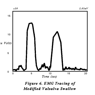
The patient should initially be educated clearly as to the nature of swallowing impairment and the goals of intervention. The therapist demonstrates the technique on his or herself with biofeedback monitoring in place. After electrodes are placed on the patient, he or she is then asked to attempt the strategy by swallowing hard and tightening the muscles in the neck and shoulders during the swallow. The patient is cued to produce high EMG peaks of short duration on the video monitor (figure 6). The treatment objective is production of increasingly higher readings. A quota for expected increase is set for each treatment session. Changes in readings are graphed to document progress.
Mendelsohn Maneuver
The superior point of entry into the esophagus is marked by the cricopharyngeal sphincter muscle (upper esophageal sphincter). In normal swallowing, this sphincter muscle remains tightly closed until the occurrence of the swallow. Vagus nerve innervation and laryngeal elevation jointly contribute to a momentary opening of the cricopharyngeal sphincter, allowing the bolus to leave the pharynx and enter the esophagus. In the case of a dysphagic patient, this muscle is impaired in its ability to maintain adequate width or duration of opening. This results in post-swallow pharyngeal residual, particularly in the pyriform sinuses. Additionally, prolonged increased pressure of the cricopharyngeal sphincter may contribute to the development of a Zenkers diverticulum immediately inferior to the muscle. The Mendelsohn Maneuver is a treatment strategy designed to facilitate cricopharyngeal sphincter opening. This strategy requires that the patient volitionally increase and maintain laryngeal elevation, thus stretching the cricopharyngeal sphincter. Since laryngeal elevation is the targeted movement, the muscles monitored are the same as those targeted for modified valsalva swallows, as mentioned above. This muscle group would specifically include the anterior belly of digastric muscle, the stylohyoid muscle and the mylohyoid muscle.
As with the modified valsalva swallow, the patient should initially receive a thorough explanation and demonstration of the treatment strategy. After electrode placement, the patient is instructed to identify the height of laryngeal elevation during the swallow and hold that position for several seconds before releasing muscular control and completing the swallowing cycle. The biofeedback line will shape boxes when the strategy is completed accurately (figure 7). The feedback line elevates off baseline with laryngeal elevation for initiation of the swallow, is maintained off the line while holding laryngeal elevation, then returns to the baseline with completion of the swallow. Treatment objectives include progressively increasing both the amplitude of the reading and the duration of elevation of the feedback line off baseline.

Relaxation of Pharyngeal Musculature
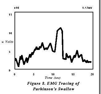
Several degenerative neuromuscular diseases result in dysphagic symptoms that require relaxation of pharyngeal musculature. In particular, Parkinsons Disease frequently results in a poorly coordinated, hyper-reactive response of pharyngeal contraction. A patient with this type of diagnosis may demonstrate adequate strength of contraction, but attempts to initiate the swallow results in a characteristic groping behavior. The patient may attempt laryngeal elevation for the swallow numerous times before actual onset and completion of the swallow. Thus, when a swallow is ultimately initiated, the effect is a weakened effort secondary to previously expended energy (figure 8). The result is an inefficient swallow with an increased risk for post swallow pharyngeal residual and malnutrition secondary to fatigue. In this case the muscles targeted are those responsible for laryngeal elevation (the suprahyoid muscle group). The target behavior is relaxation, rather than contraction, except during the occurrence of the swallow.
The objective of relaxing the pharyngeal musculature before and after the occurrence of a swallow is explained to the patient. After surface electrodes are applied, the patient is instructed to relax the muscles in the neck and throat until the biofeedback line is flat. The patient is then cued to swallow. The target behavior is a flat feedback line with a single peak for the swallow.
Conclusion
Several other authors have investigated the utilization of EMG in dysphagia diagnosis(6,7,8,12,13,15). These studies consist primarily of the application of subcutaneous needle electrode myography in the diagnosis of dysphagia and the scientific documentation of normal swallowing behavior. Further research in these areas is essential to the development of refined protocols for EMG applications. Subcutaneous electrode placement is generally not feasible or recommended for daily clinical practice; however, these studies can provide valuable information for the practicing clinician in refining surface electrode EMG biofeedback.
EMG biofeedback holds great potential as an adjunct to standard therapy for the rehabilitation of the dysphagic patient when under the direction of a speech language pathologist with expertise in this area. Biofeedback provides several critical elements that are unavailable with standard dysphagia therapy. Video monitoring allows the patient to view the swallow as it occurs, converting a previously automatic skill of which there is generally little awareness to a visually represented task that can be consciously monitored and altered. Target behavior and goal identification is measurable, providing the patient with invaluable insight into progress and motivation for continued treatment. And finally, quantitative data are produced to document the patients progress, facilitating reimbursement from third party payers. Swallowing therapy techniques are used to effect a change in the physiology of the oral and pharyngeal swallow. Biofeedback, if used appropriately, is a valuable patient aid in visualizing and documenting these changes.
REFERENCES
- Bacon, MJ: Dysphagia Treatment: A Clinical Forum. Paper presented at the 1992 American Speech Language and Hearing Association Annual Convention, Atlanta GA. 1992.
- Baker, BM, Fraser, AM, and Baker, CD: Long-Term Postoperative Dysphagia in Oral/Pharyngeal Surgery Patients: Subjects Perceptions vs. Videofluoroscopic Observations. Dysphagia 6:11-16, 1991.
- Blitzer, A: Approaches to the patient with Aspiration and Swallowing Disabilities. Dysphagia 5:129-137, 1990.
- Bryant, M: Biofeedback in the Treatment of a Selected Dysphagic Patient. Dysphagia 6:140-144, 1991.
- Buccholz, D: Neurologic Causes of Dysphagia. Dysphagia 1:152-156, 1987.
- Curtis, D, Braham, SL, Karr, S, Holborow, G and Worman D: Identification of Unopposed Intact Muscle Pair Actions Affecting Swallowing: Potential for Rehabilitation. Dysphagia 3:57-64, 1988.
- Elidan, J, Shochina, M, Gonen, B and Gay, I: Manometry and Electromyography of the Pharyngeal Muscles in Patients with Dysphagia. Arch Otolaryngol Head Neck Surg 116:910-913, 1990.
- Lidan, J, Shochina, M, Gonen, B and Gay, I: Electromyography of the Inferior Constrictor and Cricopharyngeal Muscles During Swallowing. Ann Otol Rhinol Laryngol 99:466-469, 1990.
- Lazarus, K, and Logemann, JA: Swallowing Disorders in Closed Head Trauma Patients. Arch Phys Med Rehabil 68:79-84, 1987.
- Logemann, JA: Treatment for Aspiration Related to Dysphagia: An Overview. Dysphagia 1:34-38, 1986.
- Logemann, JA: Management of the Patient with Disordered Oral Feeding. In: Logemann, JA, Evaluation and Treatment of Swallowing Disorders. College Hill Press, Inc. San Diego, 1983.
- Palmer, JB, Tanaka, E and Siebens, AA: Electromyography of the Pharyngeal Musculature: Technical Considerations. Arch Phys Med Rehabil 70:283-287, 1989.
- Tanaka, E, Palmer, J and Siebens, A: Bipolar Suction Electrodes for Pharyngeal Electromyography. Dysphagia 1:39-40, 1986.
- Tripp, F and Cordero, O: Dysphagia and Nutrition in the Acute Care Geriatric Patient. Top Clin Nutr 6(2):60-69, 1991.
- Van Overbeek, JJ, Wit, HP, Paping, RH, and Segenhout, HM: Simultaneous Manometry and Electromyography in the Pharyngoesophageal Segment. Laryngoscope 95:582-584, 1985.
- Veis, SL, and Logemann, JA: Swallowing Disorders in Persons with Cerebrovascular Accident. Arch Phy Med Rehabil 66:372-374, 1985.
Reprinted from The Biofeedback Foundation of Europe
Authors Bio:
Maggie Lee Huckabee, M.A., CCC/SLP
Senior Speech Language Pathologist
Dysphagia Specialist Massachusetts General Hospital Boston, MA