


Although on the average, vulvovaginal pain patients have less contractile capacity, It was found that there is a subset of patients who were at treatment onset, not only very tense and unstable at rest but, despite this, were also quite strong (above 25mv on initial contractile evaluation). This population posed a challenge as the tradition in pelvic floor rehabilitation was to teach the patient to exclusively localize their contractions in the pubococcygeus muscle, to the exclusion of surrounding accessory muscle. Using this training procedure, it was simply not possible to produce a contraction of adequate amplitude to reliably release the resting tension. Dr. Glazer found that permitting these patients to use the naturally occurring contractions of internal obturator, lower abdominals and adductor longus muscles, supported and enhanced the amplitude of the pelvic floor contraction adequately to "break" the resting tension level. Thus the "Glazer" protocols require the individualized "testing" of the patient with different positions and the use of different combinations of accessory muscles which enhance, rather than interfere with, the correct use of the pelvic floor muscles. This procedure is briefly outlined in a paper entitled "Functional Rehabilitation of Pelvic Floor Muscles: A Challenge to Tradition" (Glazer & MacConkey, 1996) (6).
In a 1997 paper (White, Jantos and Glazer) (7), a cohort of 32 vulvovaginal pain patients were compared to a matched control group of normals and found several sEMG characteristics which reliably differentiated the two groups. Cutoffs for these sEMG characteristics were developed and are summarized as follows:
This paper reports on a case history of a patient with classic vulvodynia who produced an sEMG protocol well within normal limits. Upon return to the gynecologist for further medical evaluation, the patient was diagnosed with cytolytic vaginosis, treated and was free of discomfort within a few days. The authors conclude, in this paper, that sEMG may be the first objective methodology for forming a differential diagnosis between functional vulvovaginal pain syndromes and other non-diagnosed medical sources of vulvovaginal pain such as infections.
Several publications are now in preparation including validating pelvic floor muscle assessment by digital palpation using sEMG as the standard (8). This study shows that sEMG is more reliable and more predictive of pelvic floor dysfunction than is digital evaluation of pelvic floor function. Another study in preparation compares sEMG biofeedback, or cognitive behavioral pain management, to vestibulectomy in the treatment of pure VVS (9). Early results suggest that biofeedback efficacy is close to that of surgery and improves over time. Research is now underway on the pattern of sEMG changes observed over treatment with focus on power density spectral frequency analysis, rise and recovery times and coefficients of variability for rest and contraction periods.
Protocol
The "Glazer" protocol for pelvic floor muscle evaluation uses a five-segment evaluation sequence as follows:
- One minute rest, pre baseline.
- Five rapid contractions (Flicks) with 10 seconds rest between each (phasic).
- Five 10 second contractions with 10 second rests between each (tonic).
- A single endurance contraction of 60 seconds (endurance).
- One minute rest, post baseline.
This protocol is a similar sequence to that used in
assessing pelvic floor muscles for incontinence. The
difference is not in the sequence of muscle actions but
the measurements taken. As mentioned earlier, the major
goal, in treating incontinence, is increasing contractile
amplitude to enhance external urethral sphincter closure
pressures. For pelvic pain, we have found that amplitude
changes are only a small part of the picture. In the
"Glazer" protocol, for each contraction and
relaxation period, integrated sEMG amplitude and standard
deviation is measured. In addition, coefficients of
variability (standard deviation divided by amplitude) are
taken as measures of muscle stability, rise and recovery
times are taken at initiation and termination of each
contraction and spectral frequencies (either FFT's or
zero crossings) are taken for each contraction. Another
difference between the "Glazer" protocol and
previous incontinence protocols is that accessory muscles
(often monitored with a second sEMG channel on lower abdominals) are not necessarily minimized. Each patient
is assessed with the use of different combinations of
accessory muscles. This is done in order to determine the
best balance between keeping the patient's focus on the
internal "lifting" sensation and, at the same
time, maximizing the use of the muscle contraction to
result in a reduction in amplitude and variability of the
subsequent rest period. We look for an exercise position,
contraction type, contraction duration, and number of
repetitions which maximize the exercise.
All patients are started on two 20 minute exercise sessions a day, each one consisting of 60 repetitions of 10 second contractions alternated with 10 second relaxation phases. All patients are required to use home training devices and intra-vaginal sensors in the conduct of their home exercises. Patients return for office evaluations every two weeks for their second and third visits and then, monthly, for subsequent visits. The frequency of office visits is determined by the observation of sEMG changes by the clinician, and compliance of the patient in the conduct of home exercises.
Over time, with continued training, we look for increased contractile amplitudes and spectral frequencies with decreased contractile coefficients of variability and rise and recovery times. In relaxation measures, we look for reduced amplitude and reduced coefficients of variability. Amplitude changes are not enough and we have seen, as mentioned earlier, many patients showing improved contractile amplitude with reduced resting amplitude and little therapeutic benefit. We believe that the spectral frequencies, rise and recovery times and coefficients of variability are related to the predominant fiber type being recruited and the coordination of use of that fiber type. The critical combination of higher amplitude contractions with higher spectral frequency, faster rise/fall times and reduced coefficients of variability suggest a predominantly fast twitch (type II) fibers. In the presence of this phenomenon (increased fast twitch coordination), the consequence is reduced amplitude and variability during rest and a reduction of the hypertonicity and instability associated with chronic uncoordinated discharge of fast twitch fibers as seen in the resting sEMG of untreated vulvovaginal pain patients.
Conclusion
Free form observations of sEMG with or without direct pelvic muscle palpation does not comprise an adequate evaluation. Replicable protocols, applied to the patient over time are necessary to assess progress. Similarly, amplitude and standard deviation measures alone are not adequate to assess changes, spectral frequencies, rise and recovery times, coefficients of variability (amplitude corrected standard deviations) must all be observed to assure that correct rehabilitation of the pelvic floor muscle is taking place. For those trained in the traditions of incontinence, it is also important to remember that you must explore positions, use of accessories, contraction duration, and number of repetitions to best achieve the desired sEMG changes. While some patients will do better with exclusive pelvic floor contractions, others cannot achieve desired outcomes without the use of accessory muscles.
References
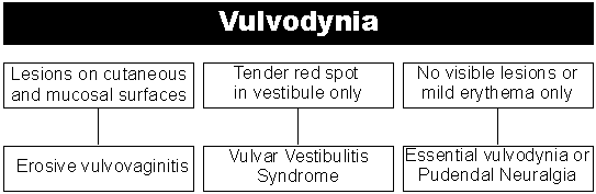
- Friedrich, E.G., Jr., Vulvar Vestibulitis Syndrome. J. Reprod. Med. 32:110-114, 1987.
- Meana, M., Binik, Y., Khalif-, S., and Cohen, D., Biopsychological Profiles of Women with Dyspareunia. Obstetrics and Gynecology 90:4:583-590.
- Marinoff, S.C., and Turner, M.L.C., Vulvar Vestibulitis Syndrome. Dermatologic Clinics 10:435-444, 1992.
- Perry, J.D. Software Standards for Perineometry, Biotechnologies, Portland, Maine, 1984.
- Glazer, H.I., Rodke, G., Swencionis, C., Hertz, R. and Young, A.W., Treatment ofVulvar Vestibulitis Syndrome with Electromyographic Biofeedback of Pelvic Floor Musculature. J. Reprod Med. 40:283-290, 1995.
- Glazer, H.I., and MacConkey, D., Functional Rehabilitation of Pelvic Floor Muscles: A Challenge to Tradition. Urol. Nurs. 16:68-69, 1996.
- White, G. Jantos, M., and Glazer, H.I., Establishing the Diagnosis of Vulvar Vestibulitis. J. Reprod. Med. 42:157-160.
- Romanzi, L., Polaneczky, M. and Glazer, H.I. A Simple Technique for Assessment of Pelvic Muscle Function as a Part of Routine Pelvic Examination; Validation by Surface Electromyography. Manuscript in preparation.
- Bergeron, S., Binik, Y.M., Khalif-, S. Pagidas, K. and Glazer, H.I. A Randomized Treatment Outcome Study of Vulvar Vestibulitis Syndrome. Manuscript preparation.
Copyright, 1997 The Biofeedback Foundation of Europe

